

Whenever I work with clients feeling mentally depleted or stuck in a state of chronic overwhelm, one of the first questions I ask is deceptively simple: When was the last time you let your body move freely through space without overthinking it? Over my twelve years as a recreational therapist, I’ve seen countless wellness trends come and go, but few things provide as immediate and tangible an emotional reset as hopping on a bicycle.
Outside of clinical programming, some of my most grounding moments happen on weekend camping trips, where packing my bike along with my tent and camera gear is non-negotiable. There is a distinct, almost immediate physical sensation that occurs roughly ten minutes into a ride: the tension behind your temples eases, your breath settles into a rhythm, and the mental fog that often clouds a hectic workweek begins to lift. In therapy sessions, we talk a lot about accessible leisure—finding movement that feels less like a punishing chore and more like genuine play.
Cycling fits this definition perfectly. It demands just enough attention to keep you anchored in the present, while your cardiovascular system quietly does the heavy lifting of sending oxygen-rich blood directly to your brain, setting the stage for emotional clarity and renewed mental energy.
Cycling is an incredible way to boost your brain’s oxygen supply while enhancing emotional resilience. As you pedal, increased blood flow sharpens your mental clarity, improving focus and memory. The endorphin rush helps relieve stress and elevate your mood, fostering emotional well-being. Plus, the community aspect of cycling nurtures connections and support. By making cycling a regular part of your life, you can transform both your mind and emotions. Discover how cycling can redefine your daily routine.
KEY TAKEAWAYS
- Cycling enhances brain oxygenation, improving focus, memory, and cognitive function through increased blood flow and oxygen delivery to the brain.
- The release of endorphins during cycling promotes mood enhancement and stress relief, contributing to emotional well-being and resilience.
- Regular cycling fosters emotional strength by encouraging individuals to confront challenges, build stamina, and push past personal limits.
- Engaging in cycling communities provides social support, fostering connections that enhance emotional resilience and motivation.
- Setting and achieving cycling goals boosts confidence and commitment, reinforcing a positive mindset and emotional clarity.
HOW CYCLING BOOSTS YOUR BRAIN’S OXYGEN SUPPLY
When you hop on your bike, you’re not just enjoying the scenery; you’re also setting in motion a powerful mechanism that boosts your brain’s oxygen supply. As you pedal, your muscles demand more oxygen, prompting your heart to pump harder. This enhanced oxygen transport elevates the flow of rich, oxygenated blood to your brain, essential for ideal functioning.
Cycling physiology plays a significant role here; it efficiently increases your lung capacity and arterial health, ensuring that oxygen reaches your brain swiftly.
With each ride, you’re not just exercising your body; you’re enhancing your cognitive abilities. The increased oxygen levels help sharpen your focus, improve memory, and even foster creativity.
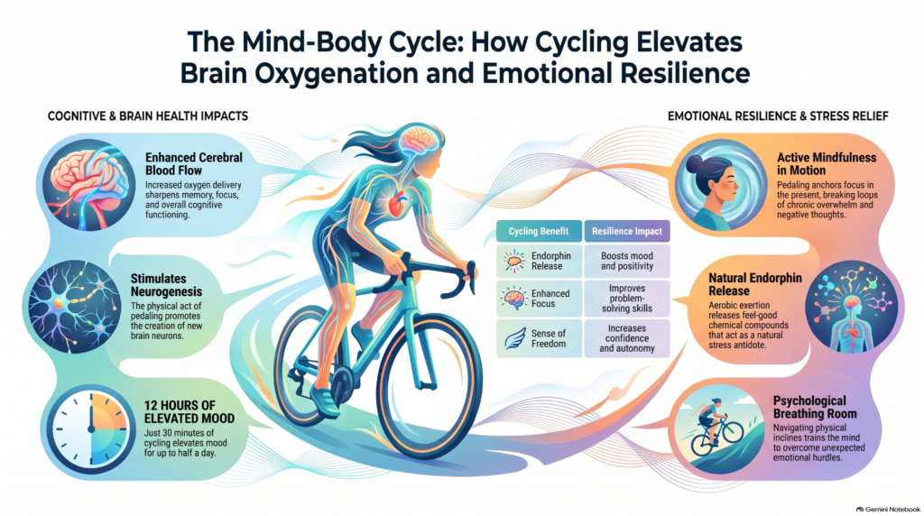
How Increased Blood Flow Enhances Mental Clarity
Exercise is known to improve cognitive functioning and the cardiorespiratory hypothesis suggests that this is due to the relationship between cardiorespiratory fitness (CRF) level and cerebral oxygenation. The purpose of this systematic review is to consolidate findings from functional near-infrared spectroscopy (fNIRS) studies that examined the effect of CRF level on cerebral oxygenation during exercise and cognitive tasks.
Increased blood flow from cycling doesn’t just fuel your muscles; it also sharpens your mind.
As your heart pumps more oxygen-rich blood to the brain, you experience enhanced cognitive function and improved mood regulation.
This boost in circulation can lead to clearer thoughts and a more focused mindset, making each ride a powerful tool for mental clarity.
Enhanced Cognitive Function
As you pedal through your cycling routine, the surge of blood flow to your brain can considerably boost your cognitive function.
This enhanced blood circulation delivers essential oxygen and nutrients, which are crucial for maintaining peak brain health. You’ll likely notice sharper focus, improved memory recall, and heightened problem-solving abilities as you ride.
The physical act of cycling promotes neurogenesis, the creation of new neurons, facilitating cognitive enhancement. With each stroke of the pedals, you’re not just exercising your body but also invigorating your mind.
The freedom of cycling allows you to escape distractions and immerse yourself in the moment, further sharpening your mental clarity.
Improved Mood Regulation
Cycling doesn’t just elevate your heart rate; it also plays a significant role in mood regulation. As you pedal, increased blood flow enhances oxygen delivery to your brain, which can help smooth out those pesky mood swings.
This surge of oxygen not only boosts your energy but also fosters emotional stability, making it easier to navigate life’s ups and downs. Engaging in regular cycling can create a sense of freedom, allowing you to break the cycle of negative thoughts and stress.
With each ride, you’re not just improving your physical health; you’re also investing in your mental clarity and resilience. So hop on your bike, feel that rush, and embrace the emotional balance that comes with it.
HOW CYCLING RELIEVES STRESS AND BOOSTS MOOD
In recreational therapy, we often pair physical movement with mindfulness principles, and cycling is essentially active mindfulness in disguise. Learning complex movement patterns—whether it’s stepping through a tricky salsa turn on a Friday evening or navigating a narrow gravel trail—forces your mind to quiet the background noise of life. When you’re on two wheels, your brain doesn’t have the bandwidth to cycle through your endless to-do lists, unresolved emails, or social anxieties; it is fully occupied with cadence, balance, the curve of the road ahead, and the breeze against your skin.
From a neurological and physiological standpoint, that steady, aerobic pedal stroke floods your cerebral tissues with fresh oxygen and naturally releases mood-elevating compounds like endorphins and endocannabinoids. I’ve watched clients who arrived at sessions visibly guarded and tense gradually open up, smile, and express a genuine sense of relief simply after twenty minutes on a scenic trail.
That rhythmic pedaling creates psychological breathing room. Over time, that physical practice translates directly into emotional resilience. You learn that when an incline gets steep and your legs burn, you don’t panic—you downshift, adjust your breathing, and trust your momentum to carry you through. That exact mindset is what helps us navigate life’s unexpected emotional uphill battles off the bike.
When you hop on your bike, you’re not just getting exercise; you’re also triggering the release of endorphins, which can greatly improve your mood.
This rhythmic movement encourages mindfulness, allowing you to focus on the present moment and escape daily stressors.
As you pedal, you’re not only enhancing your physical health but also nurturing your mental well-being.
Enhanced Endorphin Release
Engaging in regular cycling not only strengthens your body but also releases a wave of endorphins that can greatly enhance your mood and alleviate stress.
This endorphin release is one of the most significant cycling benefits, acting like a natural antidote to anxiety and depression. As you pedal through the fresh air, your brain produces these feel-good hormones, creating a sense of euphoria often referred to as the “runner’s high.”
You’ll find that your worries fade, and a sense of freedom and joy emerges. This positive emotional shift not only improves your overall well-being but also equips you to handle life’s challenges with greater resilience.
Mindfulness Through Movement
As you navigate the open road or a winding trail, the rhythmic motion of pedaling can transform your mental state, ushering in a sense of mindfulness that’s often hard to achieve in daily life.
Mindful cycling acts as a form of movement meditation, where each stroke of the pedal connects you to the present moment. The fresh air invigorates your senses, allowing stress to dissipate as you focus on the sights and sounds around you.
This practice not only relieves tension but also boosts your mood, promoting emotional resilience, much like the connection between casual gaming and mood regulation, which highlights how recreational activities can help manage stress and support emotional well-being. As you embrace the freedom of cycling, you’ll find clarity and balance, making it easier to tackle life’s challenges with newfound energy and optimism.
Emotional Benefits of Cycling Regularly
Cycling regularly not only strengthens your body but also offers significant emotional benefits that can enhance your overall well-being. As you pedal through scenic routes, you often experience a rush of endorphins, lifting your mood and reducing stress. This natural high fosters joyful connections with fellow cyclists, creating a sense of belonging within the cycling community.
Whether you ride solo or with friends, the shared passion for cycling cultivates camaraderie, encouraging you to support one another emotionally.
Moreover, cycling allows you to escape daily stresses, providing a mental break you might crave. Each ride becomes a journey of self-discovery, where you can reflect on your thoughts and feelings, promoting emotional clarity.
Engaging in this liberating activity not only nurtures your physical health but also strengthens your emotional landscape, making cycling a rewarding self care activity that helps you navigate life’s ups and downs with a more resilient spirit.
How Cycling Builds Emotional Resilience
The emotional benefits you’ve experienced from cycling serve as a foundation for building resilience in the face of life’s challenges. When you pedal through the wind, your mind clears, allowing you to confront stress with newfound emotional strength. Cycling not only builds physical stamina but also cultivates resilience building by encouraging you to push past your limits.
| Cycling Benefits | Resilience Impact |
| Releases endorphins | Boosts mood and positivity |
| Enhances focus | Improves problem-solving skills |
| Fosters community support | Builds a network of encouragement |
| Provides a sense of freedom | Increases confidence and autonomy |
Inspirational Transformations: Personal Stories of Cycling’s Impact
Anthropologists and psychologists suggest that the evolutionary roots for the cyclist’s brain go back over two million years. When humans transitioned away from a relatively sedentary ape-like lifestyle, our hunter-gatherer ancestors began to engage in mentally complex and physically demanding tasks such as foraging for food and chasing prey. Over time, our biology adapted to respond to these increased activity levels, rewarding us for engaging in tasks that combine mental engagement with aerobic exercise.
When you hear personal stories of transformation through cycling, it’s hard not to feel inspired by the profound changes individuals experience. Each personal journey showcases how cycling can redefine lives, offering a sense of freedom that many crave.
For instance, consider the story of someone who battled anxiety; through cycling, they discovered a way to channel their feelings, turning fear into empowerment. These transformative experiences aren’t just about physical fitness; they often lead to emotional breakthroughs and enhanced resilience.
People report feeling more connected to their surroundings, fostering a deeper appreciation for nature and community. As they pedal through challenges, they not only strengthen their bodies but also their minds.
The act of riding becomes a metaphor for overcoming life’s obstacles—each mile symbolizes progress and growth. These narratives remind us that cycling isn’t merely a hobby; it’s a powerful tool for personal transformation, much like journaling as recreational self-care scientific benefits that highlight how recreational activities can support emotional well-being and personal growth.
Tips for Making Cycling Part of Your Daily Life
Cycling is, essentially, a low-impact exercise that doesn’t just take you from A to B and show you the world but it’s also great for your body and mind. Its benefits include strengthening multiple muscle groups as well as improving heart health, lung capacity, and overall fitness. This sport can deliver you immense cardiovascular benefits without the high stress on your joints that occurs during high impact activities.
Incorporating cycling into your daily routine can be a game changer for both your physical and mental well-being. To make cycling a constant in your life, start small. Choose a nearby destination, like a café or park, and ride there instead of driving. This turns a mundane task into an enjoyable cycling routine.
If you face daily challenges like a busy schedule, try waking up 20 minutes earlier for a quick ride or use your lunch break to explore your surroundings on two wheels.
Consider joining a local cycling group to stay motivated and meet like-minded individuals who share your passion for freedom. Set achievable goals, whether it’s cycling a certain distance each week or participating in community rides.

RELATED STUDIES ABOUT CYCLING RECREATION AND BRAIN OXYGENATION
Incorporating cycling into your routine can be like planting a garden for your mind. Just as nurturing plants leads to vibrant blooms, cycling nurtures your brain, enhancing oxygen flow and emotional resilience. A study found that just 30 minutes of cycling can increase your mood for up to 12 hours. So, hop on your bike and cultivate that mental garden. With each pedal stroke, you’re not only boosting your clarity but also growing a more resilient, happier you.
Brain resting state functional connectivity changes with aerobic exercise, and mindfulness: A narrative review
This narrative review synthesizes empirical evidence evaluating how structured behavioral interventions—specifically aerobic exercise, mindfulness meditation, and mindful movement—alter resting-state functional connectivity (rsFC) in non-clinical adult populations, critically examining the methodological factors that explain divergent neuroimaging outcomes.
Study Overview & Conceptual Framework
- Aging and Brain Network Connectivity: Normal aging is characterized by altered neural coordination across large-scale functional brain networks. In resting-state fMRI, aging typically involves reduced within-network connectivity alongside altered between-network coupling, decreased modularity, and reduced network segregation.
- Target Resting-State Networks: Neuroimaging investigations primarily examine networks supporting higher-order cognition, memory, and emotional regulation:
- Default Mode Network (DMN): Central to self-referential processing, autobiographical memory, and resting cognition.
- Executive Control Network (ECN / Frontoparietal Network): Subserves goal-directed attention, working memory, and cognitive task execution.
- Salience Network (SAL): Integrates internal homeostatic signals with salient external stimuli to coordinate emotional and cognitive responses.
- Behavioral Interventions Across a Mind-Body Continuum: Interventions are conceptualized along an activity spectrum:
- Aerobic Exercise: Aims to increase cardiorespiratory fitness (CRF; VO2max) or moderate-to-vigorous physical activity (MVPA).
- Mindful Movement: Hybrid physical-contemplative practices (e.g., Tai Chi, Baduanjin/Qigong) combining moderate physical activity with meditative awareness and breath control.
- Mindfulness Meditation: Contemplative mental training largely operationalized through protocolized models like Mindfulness-Based Stress Reduction (MBSR).
- Research Rationale & Critical Gap: While earlier reviews emphasized positive neuroplastic remodeling from exercise and meditation, they largely overlooked variability in fMRI acquisition parameters (e.g., scan duration, motion censoring, global signal regression). This review evaluates whether reported network changes withstand methodological scrutiny.
Methodology & Search Architecture
- Search Strategy & Scope: Literature indexed in PubMed and Google Scholar published through December 2023.
- Inclusion & Exclusion Criteria:
- Multi-day interventions in non-clinical adult populations (18 years).
- Exclusively evaluated aerobic exercise, mindfulness, or mindful movement (excluding interventions combining simultaneous cognitive training during exercise).
- Excluded studies where yoga was the sole mindful movement intervention.
- Required pre- and post-intervention rsFC fMRI assessments alongside a control group.
- Study Yield: From 196 initially identified articles, 30 published papers met all inclusion criteria, representing 23 unique study populations (with several parent trials yielding multiple publications: e.g., 1 population generating 4 papers, and two separate populations generating 3 papers each).
- Intervention Breakdown: 13 exercise interventions, 11 mindfulness interventions, and 8 mindful movement interventions (with some studies evaluating both exercise and mindfulness arms).
- Sample & Scanning Characteristics Across Studies:
- Demographics: Populations averaged 55.2 years of age (median 65.5 years in intervention arms), with 16 of the 30 studies focusing specifically on older adults (>60 years). Samples were predominantly female (62% 21%) and highly educated (mean education = 14.6 to 14.9 years).
- Small Sample Sizes: Sample sizes were generally small, with mean group sizes of 29.9 (median 19.5) for intervention groups and 24.7 (median 19) for control groups. Only 5 of the 23 unique study populations (~25% of reviewed papers) included more than 30 participants per study arm.
- Scan Duration: Total scan time averaged 8.4 minutes (median 6.36 minutes; range 5.0 to 26.9 minutes). Only 7 studies met or exceeded the 9-to-10-minute scan duration recommended to achieve stable test-retest reliability.
- fMRI Preprocessing & Analysis: 21 studies applied head-motion exclusion criteria (13 person-wise, 2 run-wise, 6 using both), yielding an aggregate participant motion exclusion rate of 6.4%. Half of the studies (11/22 reporting) incorporated global signal regression (GSR). Analytic strategies varied widely, led by voxel-based seed analysis (n=9), seed-to-voxel ROI analysis (n=9), and ROI-to-ROI networks (n=6).
Key Findings by Intervention Modality
- Aerobic Exercise Interventions:
- Nine of the 13 exercise studies reported a statistically significant group time interaction favoring the exercise arm.
- The most consistent reported changes involved increased rsFC within and between nodes of the Default Mode Network and the Executive Control Network, aligning with animal models showing exercise-induced neurogenesis, angiogenesis, and elevated brain-derived neurotrophic factor (BDNF).
- However, longer exercise trials were paradoxically less likely to show broad interaction effects, and exercise dose/intensity tracking (e.g., target heart rates, MET hours) was rarely reported.
- Mindfulness Meditation Interventions:
- Ten of the 11 mindfulness studies reported significant group time interactions, with most protocols utilizing standard 8-week MBSR frameworks.
- Studies generally observed increased rsFC in the DMN and ECN alongside decreased connectivity in the Salience Network (SAL) and Motor Network (MOT).
- These shifts are hypothesized to reflect improved attentional control and the downregulation of habitual, maladaptive rumination and self-referential emotional reactivity.
- Mindful Movement (Tai Chi & Baduanjin):
- Only three published studies evaluated formal group time interaction analyses, all derived from a single underlying study population.
- Findings were contradictory—one study observed rsFC increases, another found concurrent decreases in controls, and a third noted simultaneous connectivity increases and decreases across different sub-regions of the DMN.
- Due to limited independent datasets and small cohorts, evidence regarding non-yoga mindful movement remains inconclusive.
- The Rigor Paradox (Null Findings in Well-Powered Trials):
- While smaller studies frequently reported positive intervention effects, the largest and most methodologically rigorous randomized trial reviewed (Snyder et al., 2021; utilizing large samples, multi-network parcellation, and extended scan durations) failed to detect meaningful rsFC changes following either aerobic exercise or mindfulness training.
- This discrepancy suggests that positive findings from smaller, shorter-scan studies may be inflated by low statistical power, spurious noise, or publication bias.
Methodological Vulnerabilities in the Evidence Base
- Inadequate Statistical Power: Most studies failed to report a priori power calculations. Given that stable brain-behavior and functional connectivity correlations generally require cohorts of 30 subjects per arm, the prevailing literature is prone to false positives and effect size inflation.
- Suboptimal Scan Durations: The majority of trials collected less than 8 minutes of resting-state data per session. Functional connectivity estimates derived from short scan lengths suffer from substantial intra-individual variability and low test-retest reliability.
- Network Selection Bias: The DMN was the primary ROI in the vast majority of studies. Because the DMN is the largest resting-state network, changes are statistically more probable to emerge by chance, creating a risk of false negatives in unexamined secondary networks.
- Unmonitored Physiological Confounders: None of the 30 reviewed studies assessed sleep quality or sleep duration prior to scanning sessions. Because acute sleep loss directly degrades DMN functional connectivity, unmeasured sleep differences may confound pre-to-post intervention changes.
- Sample Homogeneity & Limited Generalizability: Participants were disproportionately female (>60%), older (>60 years), well-educated (>14 years of education), and geographically restricted to the US, Europe, and China.
Recommendations for Future Neuroimaging Trials
- Enforce Rigorous Acquisition Standards: Future rsFC intervention studies should mandate resting-state fMRI runs of at least 10 minutes to stabilize functional connectivity measurements and recruit samples of at least 30 participants per condition based on formal power calculations.
- Standardize Preprocessing and Movement Censoring: Authors must explicitly report and justify choices regarding global signal regression, motion exclusion thresholds, and frame-censoring parameters to permit direct comparison across studies.
- Characterize Exercise Dosing and Fidelity: Exercise trials must quantify intervention exposure using objective physiological metrics (e.g., target heart rate zones, VO2peak, MET-hours/week) and track individual adherence rather than relying solely on attendance.
- Control for Systemic State Variables: Investigators should incorporate sleep tracking, circadian timing controls, and objective fitness verification to isolate true intervention-induced neuroplasticity from transient biological noise.
| REFERENCE: David Wing, Bart Roelands, Abraham Z. Snyder, Michael Yingling, Manuela Araque, Joshua S. Shimony, Federica Klaus, Gwen Orav, Job G. Godino, Romain Meeusen, Eric J. Lenze, Julie Wetherell, Lisa T. Eyler, Brain resting state functional connectivity changes with aerobic exercise, and mindfulness: A narrative review, Sports Medicine and Health Science, Volume 8, Issue 4, 2026, Pages 407-425, ISSN 2666-3376, https://doi.org/10.1016/j.smhs.2025.07.008. (https://www.sciencedirect.com/science/article/pii/S2666337625000800) |
Traumatic brain injury: Symptoms to systems in the 21st century
This comprehensive review synthesizes the systemic pathophysiology of traumatic brain injury (TBI), examines the underlying methodological and pharmacological factors responsible for decades of failed clinical drug translation, and proposes a 21st-century systems-based paradigm focused on early multi-target neuroprotection, whole-body stabilization, and small-volume fluid resuscitation.
Epidemiological Context & Conceptual Framing
- Global Disease Burden: TBI affects approximately 69 million individuals annually (~8,000 cases per hour) and remains the leading cause of post-injury death and permanent disability globally. While adult males exhibit nearly twice the hospitalization rate and three times the mortality of females, hospitalized females with moderate-to-severe TBI suffer significantly higher in-hospital mortality risks.
- Classification & Chronicity: Standard clinical staging relies on the Glasgow Coma Scale (GCS; mild: 13–15, moderate: 9–12, severe: 3–8). However, moderate-to-severe TBI carries a 25% to 30% acute mortality rate, and fewer than 50% of survivors regain independent functional productivity. Given long-term risks of neurodegeneration, chronic psychological disorders, cardiovascular diseases, and premature mortality, the review aligns with the 2022 Lancet Commission recommendation that moderate-to-severe TBI must be conceptualized and managed as a lifelong, chronic disease rather than an isolated, acute incident.
- Translational Impasse: Despite over 28,000 published studies and more than 250 clinical trials conducted in the past five years alone, no neuroprotective pharmacological therapy has successfully reduced human mortality or morbidity. The authors identify two primary drivers for this clinical failure:
- Widespread reliance on specific-pathogen-free (SPF) animal models in preclinical testing.
- The reductionist practice of single-nodal targeting in drug design.
Systemic Pathophysiology: The “Ripples in a Pond” Model
Primary mechanical damage (shearing, tearing, stretching of parenchyma and vessels) triggers secondary injury cascades that radiate locally and systemically through neurohormonal, vascular, and immune pathways:
- DAMPs, PRRs, and Immunometabolic Reprogramming: Within minutes of trauma, necrotic and damaged neural cells release a barrage of damage-associated molecular patterns (DAMPs; e.g., HMGB1, ATP, S100 proteins, histones, mitochondrial DNA, syndecan-1). These ligands engage pattern recognition receptors (PRRs; such as TLRs) across resident microglia, astrocytes, and infiltrating peripheral leukocytes. This receptor engagement drives a metabolic switch from oxidative phosphorylation toward aerobic glycolysis, fueling a non-resolving cytokine storm (IL-1, TNF-, IL-6) and inflammasome activation that exacerbates blood-brain barrier (BBB) breakdown.
- Sympathoadrenal Surge & Brain-Organ Uncoupling: Brain-derived inflammation triggers intense activation of the hypothalamic-pituitary-adrenal (HPA) axis and the nucleus tractus solitarius (NTS), precipitating a massive, prolonged release of catecholamines. This persistent sympathetic hyperactivation leads to the progressive uncoupling of brain-organ axes and drives multiple organ dysfunction syndrome (MODS), which occurs early in up to 68% of severe TBI patients:
- Brain-Heart Axis: Produces neurogenic myocardial stunning, ventricular wall-motion abnormalities, elevated troponin I, QT prolongation, arrhythmias, and Takotsubo cardiomyopathy.
- Brain-Lung Axis: Drives neurogenic pulmonary edema; up to 50% of ventilated severe TBI patients develop acute lung injury (ALI) or acute respiratory distress syndrome (ARDS), and ~55% acquire pulmonary infections.
- Brain-Gut Axis: Decreased mesenteric blood flow induces gut ischemia, mucosal barrier breakdown, bacterial translocation, and systemic endotoxin dissemination.
- Brain-Kidney & Brain-Liver Axes: Induces renal vasoconstriction leading to acute kidney injury (AKI in up to 20% of patients), alongside hepatic acute-phase protein release that further magnifies systemic inflammation.
- TBI-Induced Endotheliopathy & Glycocalyx Shedding: Systemic catecholamines, inflammatory cytokines, and reactive oxygen species stimulate membrane-bound sheddases that degrade the endothelial glycocalyx (a vascular protective barrier spanning >55,000m2). This shedding exposes endothelial surfaces, releases circulating biomarkers (syndecan-1, hyaluronan, thrombomodulin), impairs microvascular autoregulation, and drives localized intravascular microthrombosis through elongated von Willebrand factor (vWF)-platelet strings.
- Trauma-Induced Coagulopathy Phenotypes: Coagulopathy affects 30% to 50% of moderate-to-severe TBI cases and is linked to a ~10-fold increase in mortality risk. Unlike the hyperfibrinolytic profile typical of hemorrhagic shock, viscoelastic testing (TEG/ROTEM) demonstrates that the dominant TBI coagulopathy phenotype is a hypocoagulopathy characterized by minimal or shut-down fibrinolysis, accompanied by platelet dysfunction and consumptive hypofibrinogenemia. The review cautions that true disseminated intravascular coagulation (DIC) with widespread microvascular fibrin deposition is rare, and misdiagnosing hypocoagulopathy as DIC using routine lab scoring masks the true bleeding phenotype.
Fluid Resuscitation Controversies & Clinical Hazards
- The Narrow Resuscitation Window: While fluid therapy is essential to stabilize cerebral perfusion pressure (CPP, target 60–70 mmHg) at an optimal mean arterial pressure (MAP ~90 mmHg), both volume overload and volume restriction carry severe clinical hazards. Aggressive fluid loading induces tissue edema, hyperchloremic acidosis, hemodilution, glycocalyx damage, and pulmonary complications. Furthermore, up to 50% of critically ill adults are fluid non-responders who experience no cardiac stroke volume benefit from fluid loading.
- Normal Saline vs. Balanced Crystalloids: Normal saline (0.9% NaCl) causes hyperchloremic metabolic acidosis and renal vasoconstriction. However, the large multicenter BaSICS trial demonstrated that balanced crystalloid solutions were associated with significantly higher 90-day mortality compared to 0.9% saline in severe TBI subgroups (31% vs. 21%, p=0.02), largely due to the slightly lower osmolarity of balanced fluids aggravating cerebral edema. The authors conclude that conventional resuscitation fluids represent consensus-driven rather than evidence-based therapies, highlighting an urgent need for optimized small-volume fluid designs.
Drivers of Preclinical Translational Failure
The review argues that neuroprotective drugs fail in clinical development because preclinical testing relies on two fundamentally flawed methodological paradigms:
- The SPF Animal Artifact: Nearly all preclinical TBI studies utilize specific-pathogen-free (SPF) rodents. Introduced in the 1960s, SPF derivation relies on antibiotic treatments and barrier isolation that systematically deplete the natural gut microbiome. Consequently, SPF rodents possess immature immune systems, abnormal microvascular reactivity, blunted inflammatory responses, and divergent bleeding profiles compared to wild-type animals or humans. When exposed to surgical trauma or anesthesia, SPF animals fail to replicate normal mammalian physiology. The authors recommend that preclinical research prioritize conventionally housed, microbially intact animals, or mandate that SPF status be disclosed in Data Availability statements.
- Single-Nodal Targeting: Modern drug discovery has remained rooted in extreme reductionism, attempting to arrest secondary injury by inhibiting single molecular targets (e.g., individual cytokines like IL-1, isolated transcription factors such as NF-B, specific calcium channels, or individual free-radical pathways). Because secondary TBI is a highly redundant, multi-organ systems failure, blocking a single pathway causes uncoordinated downstream effects while failing to halt overall systemic decompensation.
A 21st-Century Systems Strategy & ALM Therapy
The authors propose shifting TBI pharmacotherapy from symptom-directed interventions toward whole-body, multi-target systems stabilization. An ideal systems-acting drug must concurrently:
- Dampen the central sympathoadrenal catecholamine storm.
- Shift early pro-inflammatory signaling to anti-inflammatory, resolving pathways.
- Protect the endothelial glycocalyx from degradation.
- Correct coagulopathy and preserve platelet reactivity.
- Maintain tissue oxygen extraction and mitochondrial ATP generation at lower perfusion pressures.
- The ALM Fluid Therapy Model: The authors have developed an intravenous small-volume resuscitation therapy combining Adenosine, Lidocaine, and Magnesium (Mg2+) (ALM):
- Physiological Mechanisms: Adenosine lowers vascular resistance, blunts inflammation, and promotes anti-ischemic metabolic arrest; lidocaine blocks aberrant sodium channels, reduces excitotoxicity, and dampens neurogenic sympathetic discharge; and magnesium stabilizes membrane potentials, blocks NMDA excitotoxic channels, and restores calcium homeostasis.
- Preclinical In Vivo Efficacy: In a rat model of moderate fluid-percussion TBI, a small IV bolus of 3% NaCl ALM followed by a 4-hour 0.9% NaCl ALM infusion achieved:
- 100% survival compared to 75% in saline controls.
- A 2.5-fold increase in cerebral blood flow at hypotensive mean arterial pressures (MAP 50–60 mmHg), maintaining parenchymal oxygen delivery while minimizing intracranial pressure elevation.
- Marked reduction in brain edema and focal lesion hemorrhage.
- Correction of post-traumatic hypocoagulopathy and preservation of platelet aggregation.
- Significant reductions in neural injury biomarkers (plasma HMGB1 down 30%, neuron-specific enolase [NSE] down 73%).
- A 70% to 80% decrease in systemic pro-inflammatory cytokines (IL-1, TNF-, RANTES).
- Full prevention of neurogenic myocardial stunning and secondary cardiac failure.
Strategic Roadmap & Research Limitations
- Preclinical Modeling Limitations: Early ALM investigations relied on anesthetized, mechanically ventilated animals observed over short acute windows (~5 hours). Future research requires unanesthetized, long-term survival models to evaluate chronic functional, cognitive, and motor recovery, alongside testing non-invasive delivery modalities (such as intranasal administration to bypass the BBB).
- Systemic Biomarker Integration: Future clinical trials must move beyond exclusive reliance on subjective GCS scores by integrating multimodal imaging with temporal biomarkers of endotheliopathy (syndecan-1), coagulopathy (viscoelastic clot-lysis profiles), and neural injury (HMGB1, NSE) to guide targeted, phenotype-specific interventions.
- Funding & Regulatory Realignment: Translating effective therapies to reduce TBI mortality requires national funding bodies and regulatory agencies to revive initiatives like the FDA’s Critical Path framework—diverting resources from isolated single-target screening toward whole-organism physiology and systems-acting therapeutics.
| REFERENCE: Geoffrey P. Dobson, Jodie L. Morris, Hayley L. Letson, Traumatic brain injury: Symptoms to systems in the 21st century, Brain Research, Volume 1845, 2024, 149271, ISSN 0006-8993, https://doi.org/10.1016/j.brainres.2024.149271. (https://www.sciencedirect.com/science/article/pii/S0006899324005250) |
Navigating Neurotoxicity and Safety Assessment of Nanocarriers for Brain Delivery: Strategies and Insights
This review article provides a comprehensive synthesis of the molecular mechanisms, physicochemical determinants, testing paradigms, and regulatory challenges associated with the neurotoxicity and safety evaluation of engineered nanocarriers designed for brain drug delivery.
Exposure and Translocation Routes to the Central Nervous System
- Primary Administration Routes: Brain drug delivery relies predominantly on parenteral (intravenous), intranasal, and oral routes. Dermal exposure remains negligible for neurotherapeutic delivery.
- Systemic Translocation via Parenteral and Oral Delivery: Intravenous administration bypasses hepatic first-pass metabolism, directly distributing nanocarriers via the systemic circulation toward the brain capillaries. Oral delivery entails translocation across the intestinal epithelium and the gut-blood barrier (GBB) into the bloodstream before reaching the cerebral microvasculature. Both systemic routes necessitate crossing the blood-brain barrier (BBB).
- Direct Olfactory and Trigeminal Translocation via Intranasal Delivery: Nasal administration allows nanocarriers deposited on the nasal mucosa to migrate along olfactory and trigeminal nerve pathways directly to the olfactory bulb and brain parenchyma, bypassing the protective endothelial tight junctions of the BBB. A fraction of inhaled/intranasally administered nanoparticles may also deposit in the respiratory tract, cross the lung-blood barrier (LBB), and access the brain indirectly via the systemic circulation.
- Alternative Brain Interfaces & Lymphatic Transport: Beyond the capillary BBB, nanocarriers interact with the choroid plexus epithelium (blood-cerebrospinal fluid [CSF] barrier) and the avascular arachnoid barrier. Nanocarriers can also traffic through deep cervical lymphatics, the cranial glymphatic network, or hitchhike within migrating immune cells (e.g., macrophages) to access brain tissue.
Molecular Mechanisms of Nanocarrier-Induced Neurotoxicity
- Cellular Adhesion, Membrane Disruption, and Tight Junction Damage: Nanocarriers adhere to endothelial and neural membranes, causing membrane perforation, loss of intracellular ATP, and necrosis. Endocytosis-mediated internalization disrupts the actin cytoskeleton, gap junction intercellular communication (degrading connexin 43 [Cx43]), and tight junction scaffolding proteins, notably reducing zonula occludens-1 (ZO-1) expression.
- Oxidative Stress & Reactive Oxygen Species (ROS) Generation: Excessive generation of ROS and reactive nitrogen species represents the primary shared mechanism of nanotoxicity. Due to high lipid content, high oxygen consumption, and weak intrinsic antioxidant defenses, the brain is exceptionally vulnerable to lipid peroxidation (elevated malondialdehyde [MDA]), protein carbonylation, and loss of antioxidant enzymes (superoxide dismutase [SOD], glutathione peroxidase [GSH-Px]).
- Neuroinflammation & Glial Activation: Nanocarriers activate resident microglia and astrocytes, triggering the release of pro-inflammatory cytokines (TNF-, IL-1, IL-6, IL-18) and inflammatory enzymes (COX-2) via MAPK, NF-B, and PI3K signaling pathways. While inorganic metallic nanoparticles are potent inflammatory inducers, organic nanocarriers (such as clodronate liposomes and uncoated PLGA nanoparticles) can also induce reactive astrogliosis, localized tissue necrosis, and microvascular hyperplastic changes.
- Mitochondrial Dysfunction: Nanoparticle accumulation prompts mitochondrial swelling, cristae disruption, loss of mitochondrial membrane potential (MMP), and impaired ATP synthesis, driving apoptotic cascades, ferroptosis, or pyroptosis.
- Genotoxicity & Endoplasmic Reticulum (ER) Stress: Direct nuclear penetration causes primary DNA strand breakage and chromosomal aberrations, whereas secondary genotoxicity is mediated by free radicals and inflammatory cascades. Overwhelming protein-folding machinery induces the unfolded protein response (UPR) and persistent ER stress, visible as structural organelle elongation and vacuolization.
- Receptor Disruption & Excitotoxicity: Nanocarriers interacting with cell-surface receptors can disrupt neurotransmission, particularly by inducing abnormal overactivation of postsynaptic NMDA receptor (NMDAR) and AMPA receptor subunits, impairing glutamate reuptake and driving excitotoxic neuronal injury.
- Glymphatic System Impairment: Accumulation of high-concentration nanomaterials within the perivascular space can impair glymphatic drainage, down-regulate aquaporin-4 (AQP-4) water channels, disrupt natural CSF outflow, and exacerbate brain edema.
Physicochemical Determinants of Neurotoxicity and Transport
- Particle Size: An inverse relationship exists between particle diameter and neurotoxicity. Smaller nanocarriers (<20–50nm) penetrate the BBB and diffuse through the 20nm brain extracellular space more readily due to high surface area-to-volume ratios, but exhibit higher surface reactivity, increased ROS generation, and accelerated cellular uptake. Particles 100nm evade rapid renal filtration and maintain longer systemic circulation, creating opportunities for delayed vascular accumulation.
- Surface Charge: Cationic (positively charged) nanocarriers exhibit higher cellular internalization rates via adsorptive-mediated transcytosis by interacting electrostatically with anionic endothelial proteoglycans. However, positive charges cause acute disruption of BBB integrity, severe hemolysis, rapid opsonization, and neuronal apoptosis. Anionic and neutral nanocarriers exhibit lower baseline uptake but offer superior biocompatibility, minimal membrane disruption, reduced protein adsorption, and safer long-term profiles.
- Shape and Aspect Ratio: High aspect ratio (AR) nanoparticles (nanorods, cylinders, filomicelles) present larger multivalent contact areas that enhance endocytic internalization compared to curved spheres. However, non-spherical shapes experience greater shear stress in blood flow, demonstrate higher cytotoxicity in astrocytes, and promote hemolysis and platelet aggregation. Spherical geometries generally offer more predictable manufacturing reproducibility and lower intrinsic tissue cytotoxicity.
- Chemical Composition & Dissolution: Organic nanosystems (polymeric nanoparticles, solid lipid nanoparticles [SLNs], nanostructured lipid carriers [NLCs], liposomes) composed of biodegradable, “generally recognized as safe” (GRAS) excipients show high biocompatibility. Conversely, inorganic nanomaterials (ZnO, Ag, CuO, TiO2, iron oxides, quantum dots) exhibit pronounced cytotoxicity due to the shedding of reactive, membrane-lytic free metal ions in aqueous biological media.
- Surface Functionalization & “Stealth” Coatings: Hydrophilic surface coatings (PEGylation, polysorbate 80, poloxamer 188) confer steric stabilization, minimize opsonization by serum proteins, reduce reticuloendothelial system (RES) clearance, and extend systemic circulation. Active targeting ligands (lactoferrin, transferrin, angiopeps, ApoE) facilitate receptor-mediated transcytosis (RMT) without compromising tight junctions. However, coatings can alter toxicity; for example, BSA- and PEG-coated silver nanoparticles elicited greater cognitive deficits and neurochemical alterations in vivo than citrate-coated formulations.
- Protein Corona Formation: Upon contact with systemic circulation, a dynamic layer of serum proteins and biomolecules rapidly coats the nanocarrier surface. This protein corona alters the nanoparticle’s synthetic identity, shifts its zeta potential, triggers agglomeration, and dictates whether it engages scavenger receptors on macrophages or target receptors at the BBB.
Neurotoxicity Evaluation Models: Capabilities & Limitations
- Physicochemical Characterization Techniques: Essential quality-control tools include Dynamic Light Scattering (DLS) for hydrodynamic diameter, Electrophoretic Light Scattering (ELS) for zeta potential, and Transmission Electron Microscopy (TEM) or Atomic Force Microscopy (AFM) for morphology and intracellular localization. Advanced optical methods, such as Real-Time All-Optical Nanoparticle Analysis by Scattering Microscopy (RONAS), enable label-free tracking of protein corona adsorption directly within full serum.
- Conventional 2D In Vitro Assays:
- Assay Types: MTT/MTS/XTT/WST-1 (mitochondrial dehydrogenase metabolic activity), LDH release and Trypan blue exclusion (membrane permeability), Annexin-V/PI (apoptosis/necrosis flow cytometry), Comet and micronucleus assays (genotoxicity), and DCFH-DA or Electron Paramagnetic Resonance (EPR; oxidative stress).
- Artifact Vulnerabilities: Nanomaterials frequently interfere with absorbance, fluorescence, and luminescence readouts, adsorb chemical reagents to their surfaces, or contain endotoxin/LPS contaminants that generate false-positive cytotoxicity data. Furthermore, static monocultures lack physiological fluid shear stress and 3D architectural complexity.
- In Vivo Animal Models:
- Species: Rodents (mice, rats), zebrafish embryos/larvae, Drosophila melanogaster, and Caenorhabditis elegans.
- Strengths & Weaknesses: In vivo platforms capture whole-organism pharmacokinetics, neurobehavioral endpoints, and multi-organ systemic cascades. However, they are resource-intensive, low-throughput, constrained by the 3Rs ethical framework, and hindered by species-specific differences in plasma constituents, cerebrovascular anatomy, and BBB transporter kinetics.
- Advanced & Microphysiological In Vitro Models:
- 3D Brain Organoids: Derived from human induced pluripotent stem cells (iPSCs), organoids replicate 3D cytoarchitecture, cell-cell signaling, and human genetic backgrounds. Limitations include lack of vascularization, nutrient-deficient necrotic cores, and batch-to-batch structural heterogeneity.
- Microfluidic Brain-on-a-Chip: Incorporates microchannels, dynamic fluid flow, and extracellular matrix scaffolds to subject co-cultured brain endothelial cells, astrocytes, and pericytes to physiological shear stress. Integrated micro-electrode arrays (MEAs) and transendothelial electrical resistance (TEER) sensors enable real-time, non-invasive electrophysiological and barrier-integrity monitoring.
- 3D and 4D Bioprinting: Uses bioinks (hydrogels) to pattern functional multi-cellular neural tissue constructs. Evolving 4D bioprinting incorporates stimuli-responsive transformations over time (responding to pH, temperature, or enzymes) to model dynamic neurodevelopmental microenvironments.
- In Silico & Computational Toxicology: Employs Quantitative Structure-Activity/Toxicity Relationships (QSAR/QSTR), molecular dynamics simulations, and machine learning models to predict membrane lipid extraction (e.g., graphene-induced destructive phospholipid extraction) and tissue-specific neurotoxic endpoints based on nanocarrier molecular descriptors.
Regulatory Landscape & Strategic Challenges
- Regulatory Fragmentation: Formal regulatory standards tailored specifically to nanomedicine neurotoxicity remain underdeveloped. While agencies like the US FDA, European Commission (NANoREG), and OECD provide broad guidelines (e.g., OECD TG 424 for rodent neurotoxicity, GD 39 for inhalation, and REACH annexes), regulatory evaluations are performed on an ad-hoc, case-by-case basis.
- Dosing Metric Ambiguities: Studies lack consensus on whether to express nanoparticle dose metrics in terms of mass concentration (mg/mL), particle number concentration, or total biologically available surface area, leading to contradictory threshold interpretations across published literature.
- Neglect of Chronic, Low-Dose Exposure: The majority of preclinical nanotoxicology focuses on high-dose acute administration, failing to model real-world clinical realities where patients with chronic neurodegenerative diseases require repetitive, low-dose maintenance regimens over years. Long-term deposition, cumulative biopersistence, and delayed cognitive effects remain under-characterized.
- Safe-by-Design and Quality-by-Design (QbD): Overcoming translational bottlenecks requires integrating toxicological screening directly into the early design phase—optimizing biodegradable matrices, selecting neutral or moderately negative surface charges, incorporating dynamic protein corona evaluations in human serum, and harmonizing multi-omics computational pipelines with organ-on-a-chip validation.
| REFERENCE: Maria Inês Teixeira, Carla M. Lopes, Maria Helena Amaral, Paulo C. Costa, Navigating Neurotoxicity and Safety Assessment of Nanocarriers for Brain Delivery: Strategies and Insights, Acta Biomaterialia, Volume 189, 2024, Pages 25-56, ISSN 1742-7061, https://doi.org/10.1016/j.actbio.2024.09.027. (https://www.sciencedirect.com/science/article/pii/S1742706124005439) |
CONCLUSION
Looking at the science connecting aerobic movement, cerebral blood flow, and emotional stability, it’s easy to get lost in clinical terms like neurogenesis and functional connectivity. But at its core, cycling isn’t about metrics, speed, or tracking data—it’s about carving out a dependable sanctuary for your mental health. In my own life and in over a decade of practice, I’ve found that true resilience isn’t built through grueling discipline; it’s cultivated through joyful, sustainable habits that consistently fill your emotional cup.
Whether you’re pedaling through a quiet neighborhood park at dawn, commuting to work, or exploring unpaved backroads on a Saturday morning, cycling offers a direct pathway to clearing mental clutter and restoring inner balance. It reminds us that our physical bodies and emotional landscapes are deeply intertwined: when you feed your brain the oxygen and movement it craves, your emotional outlook naturally follows suit.
You don’t need high-end racing gear or an intense training regimen to reap these rewards. All you need is an open mind, an accessible path, and the willingness to let two wheels carry you away from daily stress and back toward yourself.